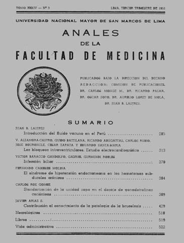
The intraventricular blocks - Electrocardiographic Study
DOI:
https://doi.org/10.15381/anales.v34i3.9528Abstract
Modifications r waves early in the right precordial leads when a left locking occurs due to abnormal excitation current in the septum. The way of activation wave progresses in the regions engaged by the lock and the abnormal orientation of the vectors representing the electric forces in these regions, may explain the QRS-T changes observed in the electrocardiograms at the left locking ensue . Analysis of correlative changes "Waves B" or waves and blocking waves represent the normal ventricular activation clarification pathogenesis of the left blocks. The left blocks are manifested electrocardiographically as "intramural". The different lock types are to be left to the extent and magnitude of functional impairment of specialized muscle fibers in which the activation proceeds very quickly. For its size the left blocks are "partial" or "total". The duration of the ventricular complex does not define whether a left block is "complete" or "incomplete". In a given shape derivation electrocardiogram can indicate whether a left block is "complete" or "incomplete". A left block is "incomplete" in the explored area when "B wave" can coexist with deflections represent normal activation of left ventricular muscle. A left block is "complete" in the explored zone when there are only "B wave" and there is no evidence of inflections representing normal activation. In the text the difficulties and limitations that exist for the electrocardiographic diagnosis of left ventricular hypertrophy are discussed.Downloads
Published
1951-09-17
Issue
Section
Trabajos originales
License
Copyright (c) 1951 V. Alzamora Castro, Guido Battilana, Ricardo Abugattas, Carlos Rubio, José Bouroncle, César Zapata, Eduardo Santa-María

This work is licensed under a Creative Commons Attribution-NonCommercial-ShareAlike 4.0 International License.
Those authors who have publications with this magazine accept the following terms:
- Authors will retain their copyrights and guarantee the journal the right of first publication of their work, which will be simultaneously subject to Creative Commons Attribution License that allows third parties to share the work as long as its author and its first publication this magazine are indicated.
- Authors may adopt other non-exclusive licensing agreements for the distribution of the version of the published work (eg, deposit it in an institutional electronic file or publish it in a monographic volume) provided that the initial publication in this magazine is indicated.
- Authors are allowed and recommended to disseminate their work over the Internet (eg: in institutional telematic archives or on their website) before and during the submission process, which It can produce interesting exchanges and increase quotes from the published work. (See El efecto del acceso abierto ).
How to Cite
1.
Alzamora Castro V, Battilana G, Abugattas R, Rubio C, Bouroncle J, Zapata C, et al. The intraventricular blocks - Electrocardiographic Study. An Fac med [Internet]. 1951 Sep. 17 [cited 2024 Jul. 6];34(3):313-69. Available from: https://revistasinvestigacion.unmsm.edu.pe/index.php/anales/article/view/9528


