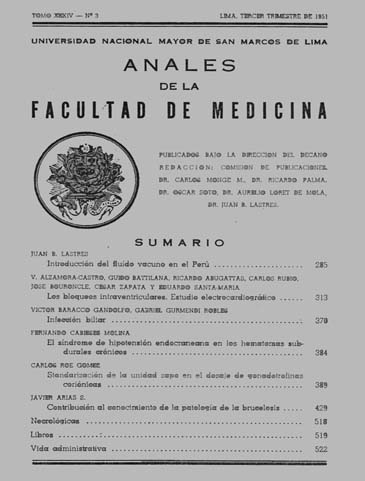
Contribution to the knowledge of the pathology of brucellosis
DOI:
https://doi.org/10.15381/anales.v34i3.9532Abstract
1. A histo-pathological study of 6 fatal cases of brucellocies is presented, in 4 out of the 6 cases a post-mortem bacteriological study was possible because spleen, lymph-nodes and liver suspensions were inoculated in guinea piqs and isolated from these animals Br. melitensis. In one case (N° 4) the aglutination test was positive with pleural exudate. In other case (N° 5) it was positive in the ascitic fluid. 2. Description of lesions due to Brucella not found in the literature, are as follows: a) Subacute atrophy of the liver; with the finding of the microorganism in samples from that organ. b) Meningo-encephalitis and meningitis due to Br. melitensis; cases with bacterilogical findings. c) In the suprarenal glands, granulomas due to Brucella. d) Brucellar salpingitis with granuloma formation; case with positive bacteriological finding. 3. It is pointed out the frequency and importance of the vascular lesions showing cases of endophlebitis in the splen and the kidney. The frequently mentioned cases of trombosis and aneurisma, in the literature, are interpreted as a result, of above mentioned vascular lesions. 4. A pre-cirrotic histological picture is found in a case of severe hepatitis of long clinical evolution. 5. It is pointed that in our enviroment the hepatic damage of median or great intensity is more frequent than in other; this is explained, in port. by the association of nutritional deficiency agraviating the injury on the liver. The following clinic-pathological types of hepatic damage, are distinguished: a) hepatitis; b) acute or subacute hepatitis; c ) cronic hepatitis (pre-cirrosis and cirrosis ): and d) acute or subacute atrophy of the liver. 6. Description of lesions due to Brucella similar those given by several authors, are the followinc : intersticial nephritis, myocarditis, peritonitis, hepatitis and granulomas in lymh-nodes, spleen and bone marrow. 7. Anatomical proofs of reactivation of old tubercle foci, that happened during the brucella infection are presented, giving an explanation of inter-relations of these processes. 8. The author arrives to the conclusion that there is not a uniform tissue reaction to the brucellosis agretion. The fundamental alterations take place in the RES, but other tissues react also. The following inflamatory reactions are distinguished: a) RES. hiperplasy; b) infiltrative-proliferative inflamation; and c) infiltrative-exudative inflamation. These different reaction can occur on the same individual. The three, can occurr as diffuse, not circunscribed infiltrate, or as true granuloma delimited foci. The more typical finding has been the granuloma formed by the necrosis of large monocite's foci . The great diversity of the anatomo-pathological picture is interpreted as dependent on the hypersensibility, resistance, virulence and number of germs; observations on the importance of each one of these factors are presented. 9. Clinical and anatomo-pathological observations on 35 guinea piqs inoculated with a strain of Br. melitensis isolated írorn ahuman autopsy are presented. Slight alteration of the blood picture are noted, as well as, the rapid appearance of positive agglutination test in animals inoculated with material from human autopsies. 10. The histological characteristics of the hypersentivity reaction in the guinea pig are described. 11. A systematic study of the reactions elucidated by intracutaneous íniectíons of Brucella melitensis in the guinea pig is given, with the description of macro and micrascopic characteristics. It has been possible to demostrate the intracelular multiplication of Brucella day by day, developing in the macraphages of inflamatory focus. Between the 9th and the 13th day after inoculation 100% of the macraphages showed the organisms. 12. The reinfection in guinea pig gives place to more rapid, acute necratic lesions, that dissapear in a shorted time, compared with first infection. 13. Clinical and anatomopathological observations in dogs infected with Br. melitensis, are shown. A very natural resistence in this animal is found. As in other infections, the splenectomy, would lower the natural resistence. 14. The study of the local action of the first infection in the dog compared with the same in guinea pigs. Shown rapid appearance of inflamatory reaction and ulceraction; early involution and not obvious multiplication of the organisms. 15. The study of intradermal and intrahepatic reinfection in dog shown more necrotic and severe lesions than in the first infection, which we think are due to a high degree of bacterial hypersensitivity. The effect of traumatisms on the regional symptomatology is shown, based on experimental observations.Downloads
Published
1951-09-17
Issue
Section
Trabajos originales
License
Copyright (c) 1951 Javier Arias S.

This work is licensed under a Creative Commons Attribution-NonCommercial-ShareAlike 4.0 International License.
Those authors who have publications with this magazine accept the following terms:
- Authors will retain their copyrights and guarantee the journal the right of first publication of their work, which will be simultaneously subject to Creative Commons Attribution License that allows third parties to share the work as long as its author and its first publication this magazine are indicated.
- Authors may adopt other non-exclusive licensing agreements for the distribution of the version of the published work (eg, deposit it in an institutional electronic file or publish it in a monographic volume) provided that the initial publication in this magazine is indicated.
- Authors are allowed and recommended to disseminate their work over the Internet (eg: in institutional telematic archives or on their website) before and during the submission process, which It can produce interesting exchanges and increase quotes from the published work. (See El efecto del acceso abierto ).
How to Cite
1.
Arias S. J. Contribution to the knowledge of the pathology of brucellosis. An Fac med [Internet]. 1951 Sep. 17 [cited 2024 Jul. 6];34(3):429-517. Available from: https://revistasinvestigacion.unmsm.edu.pe/index.php/anales/article/view/9532


