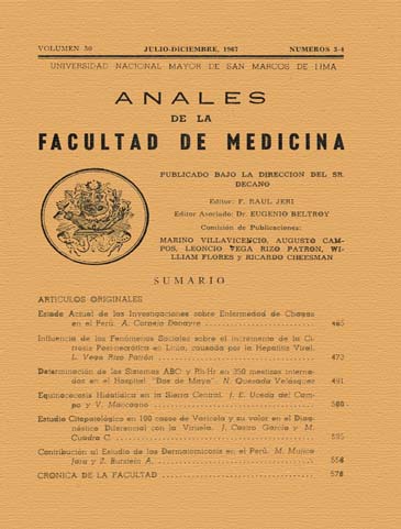
Cytopathological study of 100 cases of varicella and its value in the differential diagnosis of smallpox
DOI:
https://doi.org/10.15381/anales.v50i3-4.5547Abstract
Disclosed cytologic features of a cell type with diagnostic value in chickenpox , called Giant Cell Features , Multinucleated Giant Cells , Atypical cells or " pseudotumoral " . These cells acquire cyto logical morphologically distinguishable characteristics , dye , quantitative , and fine percentage , according to the stage of the rash variceloso are. In phase transparent vesicle are usually round or oval , with one or two giant nuclei , acidophilic staining affinity discreetly . In phase turbid or opalescent vesicle there is a tendency of irregular deformation of cells and nuclear multi-segmentation ( multinucleated ) and acquire more acidophilic staining in the previous phase. At the stage of pustule are often spherical with giant core and take an acidophilus frankly coloring with Leishman . The cytoplasm of these cells is scarce and generally takes basophilic staining in the three phases of variceloso rash. In all of them there is loss of core - cytoplasmic ratio . Almost all of these cells have a clear perinuclear halo, perfectly highlighted in the cell cytoplasm. In none of these cells has identified the nucleolus . The size of these cells is also variable ( 40 to 60 microns ) . The average standard size, average core - cell maximum size , the average core - cell minimum size are higher in the turbid vesicle phase or in phase opalescent transparent vesicle and pustule ; in this last considerably less than in the above. Quantitative assessment of the percentage of these cells relative to other also varies the phase of the varicella vesicle , being 100% in the clear vesicle , 40 to 60% in the opalescent vesicle and 2 % in the pustule . These cells have not been found in the three cases of smallpox have been studied from the point of view cytological , following the same procedure used in technical chickenpox. Pathological studies of representative complementary in chickenpox and in three cases of smallpox cases confirm the presence of these cells in the first and Guarnieri corpuscle in the second.Downloads
Published
1967-12-18
Issue
Section
Trabajos originales
License
Copyright (c) 1967 Jorge Castro García, Manuel Cuadra C.

This work is licensed under a Creative Commons Attribution-NonCommercial-ShareAlike 4.0 International License.
Those authors who have publications with this magazine accept the following terms:
- Authors will retain their copyrights and guarantee the journal the right of first publication of their work, which will be simultaneously subject to Creative Commons Attribution License that allows third parties to share the work as long as its author and its first publication this magazine are indicated.
- Authors may adopt other non-exclusive licensing agreements for the distribution of the version of the published work (eg, deposit it in an institutional electronic file or publish it in a monographic volume) provided that the initial publication in this magazine is indicated.
- Authors are allowed and recommended to disseminate their work over the Internet (eg: in institutional telematic archives or on their website) before and during the submission process, which It can produce interesting exchanges and increase quotes from the published work. (See El efecto del acceso abierto ).
How to Cite
1.
Castro García J, Cuadra C. M. Cytopathological study of 100 cases of varicella and its value in the differential diagnosis of smallpox. An Fac med [Internet]. 1967 Dec. 18 [cited 2024 Jul. 16];50(3-4):525-57. Available from: https://revistasinvestigacion.unmsm.edu.pe/index.php/anales/article/view/5547


