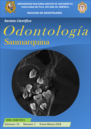
Quantitative cervical vertebral maturation assessment with the use of Cone-Beam computed tomography
DOI:
https://doi.org/10.15381/os.v21i1.14429Keywords:
Evaluation, Bone development, Cervical vertebrae, Tomography X-Ray computedAbstract
Objective: To evaluate a quantitative method of bone maturation in cervical vertebrae in 14 Cone Beam computed tomographies of children aged 9 to 15 years old obtained from the archive of the Radiologic Service of the School of Dentistry at the National University of San Marcos. Methods: A retrospective, descriptive and transversal study was carried out. The sample was for convenience of 14 tomographies of children (seven male), these were clinically justified images, previously taken with the Picasso Master 3D - FOV 20x15 and processed with the EzImplant software; the head was positioned with reference to the Frankfort plane and the cuts were made. We used a quantitative method that established four stages of maturation in an objective way by means of an equation that used three measurements in the vertebrae C2, C3 and C4. Results: The highest percentage of children was found in the deceleration period. In the high-speed period, the highest values were found for the female sex with an average age of 11 years. Conclusions: The quantitative method described was simple, practical and applicable through Cone Beam computed tomography showing good results.Downloads
Downloads
Published
Issue
Section
License
Copyright (c) 2018 Rocío del Pilar Ríos León, Jessica Salas Huallparimache, Cynthia Salazar Zapata, Sue Salas Catacora, Gina Flores Díaz, Carlos Tisnado Florián, Daniel Blanco Victorio

This work is licensed under a Creative Commons Attribution-NonCommercial-ShareAlike 4.0 International License.
AUTHORS RETAIN THEIR RIGHTS:
a. Authors retain their trade mark rights and patent, and also on any process or procedure described in the article.
b. Authors retain their right to share, copy, distribute, perform and publicly communicate their article (eg, to place their article in an institutional repository or publish it in a book), with an acknowledgment of its initial publication in the Odontología Sanmarquina.
c. Authors retain theirs right to make a subsequent publication of their work, to use the article or any part thereof (eg a compilation of his papers, lecture notes, thesis, or a book), always indicating the source of publication (the originator of the work, journal, volume, number and date).


