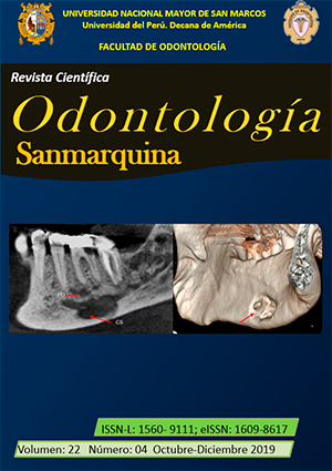
Unusual Stafne´s osseous cavity presentation: computerized tomography and magnetic resonance imaging study
DOI:
https://doi.org/10.15381/os.v22i4.17053Keywords:
Mandible, Computed tomography, Magnetic resonance imaging (source: MeSH NLM)Abstract
The Stafne bone cavity (SC) has been described as an oval radiolucence, with defined and corticalized edges located below the jaw duct between the first molar and the angle of the jaw. Atypical cases of presentation of the cavity in lobed form with irregular, sclerotic or incomplete margins, as well as an unusual location require the use of imaging methods that make possible a differential diagnosis, avoiding an invasive procedure. The purpose of this work was to describe a case of SC in a 74-year-old male patient, with a history of prostate cancer. Cone beam computed tomography images showed an open cavity toward the lingual table below the mandibular canal. Magnetic resonance imaging and multi-cut computerized tomography allowed identifying the defect content, found adipose tissue. The radiographic examination of an atypical SC should be complemented with tomographic and magnetic resonance studies; these provide relevant information to the definitive diagnosis, limiting the performance of a surgical examination. In the clinical case presented, the characterization of the extension of the defect, its relationship with neighboring teeth and structures, as well as the identification of the content allowed us to rule out the presence of a prostate cancer metastasis.
Downloads
Downloads
Published
Issue
Section
License
Copyright (c) 2019 Adalsa Hernández-Andara, Ana Isabel Ortega-Pertuz, Juan Saavedra, Marcos Gómez, Mariana Villarroel-Dorrego

This work is licensed under a Creative Commons Attribution-NonCommercial-ShareAlike 4.0 International License.
AUTHORS RETAIN THEIR RIGHTS:
a. Authors retain their trade mark rights and patent, and also on any process or procedure described in the article.
b. Authors retain their right to share, copy, distribute, perform and publicly communicate their article (eg, to place their article in an institutional repository or publish it in a book), with an acknowledgment of its initial publication in the Odontología Sanmarquina.
c. Authors retain theirs right to make a subsequent publication of their work, to use the article or any part thereof (eg a compilation of his papers, lecture notes, thesis, or a book), always indicating the source of publication (the originator of the work, journal, volume, number and date).


