
Stafne bone defect: findings on panoramic radiography
DOI:
https://doi.org/10.15381/os.v23i2.17768Keywords:
bone defectAbstract
Sr. Editor.
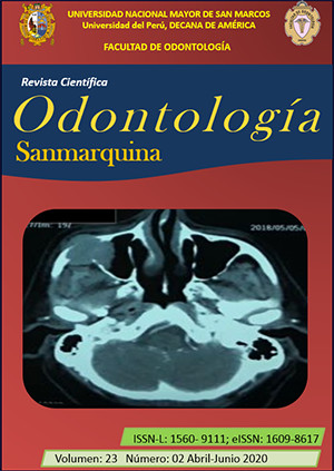
La cavidad ósea de Stafne es un defecto óseo de forma redondeada u ovalada, puede presentarse de manera unilateral o bilateral teniendo un diámetro promedio de entre 1 a 3 cm. Mayormente se encuentra ubicada en la zona posterior lingual de la mandíbula por debajo del conducto del nervio dentario inferior, pero también se puede localizar en otras zonas de la mandíbula. Esta entidad, al no generar sintomatología, generalmente es un hallazgo en la radiografía panorámica odontológica; pudiendo describirse como una imagen radiolúcida unilocular de forma redondeada, de límites definidos y bordes corticalizados (Figura) 1-6.
Downloads
Downloads
Published
Issue
Section
License
Copyright (c) 2020 Fernando Russbelts Sthorayca Retamozo, Carlos Darío Merino Bustamante, Vilma Elizabeth Ruiz García de Chacón

This work is licensed under a Creative Commons Attribution-NonCommercial-ShareAlike 4.0 International License.
AUTHORS RETAIN THEIR RIGHTS:
a. Authors retain their trade mark rights and patent, and also on any process or procedure described in the article.
b. Authors retain their right to share, copy, distribute, perform and publicly communicate their article (eg, to place their article in an institutional repository or publish it in a book), with an acknowledgment of its initial publication in the Odontología Sanmarquina.
c. Authors retain theirs right to make a subsequent publication of their work, to use the article or any part thereof (eg a compilation of his papers, lecture notes, thesis, or a book), always indicating the source of publication (the originator of the work, journal, volume, number and date).

